Sunday, April 27, 2008

Are You Eating Right For Your Blood Type?

The concern about obesity and its associated risk factors for hypertension and cardiovascular diseases is not something new to the public. Today, even cancer, leukemia, diabetes mellitus, acne, stroke and osteoporosis, which were once upon thought to be the jargons of the medical sciences, are becoming the centre of attention for people from all walks of life. Why such unprecedented blooming interest, you may ask? The simple reason being that human beings are fear of death - a issue still considered taboo to many, but a definite fact that all beings that live must endure. Perhaps death may not be an issue after all. It may be the long term or possibly a life-long physical or emotional sufferings that are the most difficult to counter. Nevertheless, with the intervention of various technologies, medical sciences are gradually progressing into a wonderful breakthrough. Many diseases primitively thought to be the play-of-the-devil are evidently proved otherwise and several terminal illnesses have been successfully treated with only simple involvement of certain tools. How about prevention-wise?

While there is still no scientific prove about the existence of an elixir of life, many regimens from different schools of thoughts are promising ‘eternal’ health with strict adherence. There are various articles about the magnificent power of evening primrose oil and that of fish oil, saw palmetto, glucosamine, coenzyme Q, etc., but what that caught my attention most was an article about eating according to the blood type. I have come across similar diet regimens and their associated risk of diseases with different astrological Sun signs, a notion I take for the mere pleasure of reading but will not indulge into it seriously since astrology is not a true medical science. But when a diet design is connected to the well-established ABO blood group system, it suddenly resonated to me again and again that I should take a quick look at this article. After all, we are what we eat. I, having known my blood type already may as well know what’s best for me. I’ll definitely do not want to eat something that’s not coded for in my genes.

The blood type diet actually revolves around the evolutionary history of how different blood groups came into existence with time and that we should adhere to the types of diet intended for that time that coincide with the blood group. Dr. Peter D’Adamo, author of the best selling book Eat Right For Your Type declares that our ancestors were all type O skilled hunters that thrived on meat based diet. They possessed genetic memory of strength and endurance from the high protein diet. By 25000 B.C. – 15000 B.C. type A blood type emerged when people started to learn the techniques of crops cultivation. These so-called cultivators emphasized a diet free of red meat with preference for vegetables and more calming exercises. As climatic changes in the western Himalayan mountains progressed, blood type B appeared. According to Dr. D’Adamo, type B are nomads with strong immune and flexible digestive systems. They are the only ones who can thrive on dairy products. Blood type AB on the other hand, is the most recently evolved type and stays intermediate between blood types A and B in terms of dietary needs. The key to optimal health here is to eat as our ancestors with the same blood type ate and engage in the same kinds of exercises they did.

Dr. D’Adamo related his research based on the effects of lectin, a type of protein found commonly in eaten foods, particularly the seeds of leguminous plants. According to him, certain lectins are incompatible with certain blood types and this incompatibility causes lectins to attract to and clump the red blood cells through a process called agglutination once they are absorbed from the digestive tract into the bloodstream. Since lectins react differently with each ABO type antigen, there is a need for a selection of different foods for O, A, B and AB blood types to minimize its reactions. Henceforth, Dr. D’Adamo divided foods into 16 categories with 3 types of labels – highly beneficial, neutral and avoid, according to each of the 4 blood types for reference.

Another interesting point is that type O people are blessed with strong stomach acid and enzymes, thus they are able to metabolize almost everything. On the other hand, if a blood type A individual who already has thick blood is consuming food incompatible with his blood type, the blood will eventually become even thicker due to the agglutination effect and hence a risk for hypertension and heart disease. This phenomenon is rarely observed in type O since they start out with the thinnest blood, and if any agglutination takes place, the blood will not thicken to the extent experienced by other blood types. Nevertheless, with improper dietary intervention, people of all blood types will eventually end up with diseases – the risk being higher for types A and AB in the short run but longer for people with types O and B. Since our blood groups were already determined at the time of conception and cannot be changed, we may have the privilege to live a longer and healthier lifestyle if we can change the way we eat.

Does this mean that a blood type O person should include meat in almost all of his meals and engage in vigorous exercises like running and cycling? How about the risk of cardiovascular diseases and renal failure with a high protein diet? Are there any clinical trials to support such claims? Unfortunately, Dr. D’Adamo’s statements lack solid scientific support with no comparative clinical trials on the efficacy of his regimen. His research on lectins was done in vito and nobody guarantees similar results physiologically in the real humans. Moreover lectins are destroyed by cooking or digestive enzymes and the amount of lectins absorbed intact through the digestive system is minimal. Furthermore, to design a diet based on the ABO blood group system alone may sound quite naive since there are more than 30 unique markers identified on the surface of red blood cells. Phylogenetic studies of human and non-human ABO alleles on the other hand showed that A gene was the first to evolve although O blood type is common in all populations around the world, another notion that disproved Dr. D’Adamo’s theory.

Nevertheless, the decision is yours whether to believe it or not. I personally feel that there are to a certain extent some controversies in the world of science as new discoveries begin to surface. For instance, we were once told that unsaturated fats are better than saturated fats and now both of them seem to be bad for health for one reason or another. From another point of view, Lingzhi and aleo vera may be a good source of remedy but they sometimes failed just because different individuals react differently with the same substance. Similarly, drug A may be efficient in alleviating your illness but it may not be a magic bullet to another patient. By the way, there’s a connection between different types of ABO antigens and the preference of certain bacteria and virus on eliciting infection. In a nutshell, I strongly believe that there’s a connection somehow, perhaps a different type of relationship not similar to that of Dr. D’Adamo’s but in terms of dietary-wise, I feel that moderation in all types of food is the key to good health. What do you think?

Saturday, April 19, 2008

ROS and Diabetic Complications

1. Reactive Oxygen Species

A free radical is any species with one or more unpaired electrons in its outer orbital, and is thus electronically unstable and tends to react with other neighbouring species to achieve a more thermodynamically stable configuration with no net spin. These radicals and other non-radicals that are themselves easily converted to free radicals are collectively termed the reactive species (Evans et al., 2002).

The molecular oxygen (O2) itself is a less reactive bi-radical with two unpaired electrons located in a parallel spin at a different π orbital. Such configuration confers a spin restriction on the reacting molecules (Yu, 1994) and eventually creates a one-electron reduction transfer that leads to the generation of ROS, notably the superoxide (O2•-), hydrogen peroxide (H2O2) and the hydroxyl radicals (•OH) (Figure 1).


ROS

In fact, ROS are continuously produced in the mitochondrial respiratory chain where O2 consumption is a necessity for all aerobic biological systems. Nevertheless, the damages inflicted by these reactive species are usually minimal due the protective roles of antioxidants in the body. However, the equilibrium of prooxidant-antioxidant pool is often breached during stress and diseases and leads eventually to a phenomenon termed oxidative stress.

2. Generation of Free Radicals in Diabetes Mellitus


Hyperglycaemia and possibly free fatty acids (FFAs) have been widely correlated with the production of free radicals that implicates in the pathogenesis of diabetes and diabetic complications (Evan et al., 2002). Blood capillary endothelial cells, pericytes, lens fibre cells and peripheral neurons that exhibit insulin-independent uptake of glucose by GLUT1, and thus a free interchange of glucose from the extracellular to the intracellular environment, are often the main targets of glucotoxicity.

2.1 Superoxide production

In a hyperglycaemic state, excess electron donors (NADH and FADH2) will be generated from the TCA cycle during the catabolism of glucose and lead to a high mitochondrial membrane potential, ∆μH+ by pumping H+ across the inner mitochondrial membrane. However, beyond a certain threshold value of ∆μH+, electron transport to complex III will be inhibited and electron is instead transferred to O2 for reduction to O2•- (Korshunov et al., 1997). The overexpression of uncoupling protein-1 (UCP-1) that dissipates ∆μH+ has been shown to prevent hyperglycaemia-induced overproduction of ROS (Nishikawa et al., 2000). Similarly, elevated FFAs has also been noted to induce uncoupling of oxidative phosphorylation that leads to the generation of more mitochondrial O2•- (Bakker et al., 2000).

The overproduction of mitochondrial O2•- is proposed to be the unifying source that diverts the propagation of oxidative stress in diabetes mellitus into various pathways of glucose overutilisation as described after forth, and hence more free radical production (Figure 2).


Glucose overutilisation
Figure 2: Alternative pathways of glucose overutilisation in diabetes mellitus that lead to the generation of ROS.

2.2 Polyol pathway

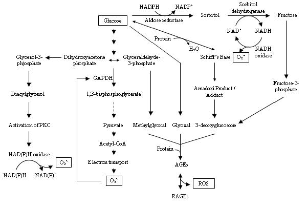
Aldose reductase is the first enzyme in the polyol pathway with low affinity for glucose. Under euglycaemic condition, the metabolism of glucose by this pathway is limited but in a hyperglycaemic state, excess glucose will result in its increased conversion to sorbitol with the concomitant decrease in NADPH (Swidan and Montgomery, 1998). The depletion of NADPH, which is also required for glutathione reductase to regenerate reduced glutathione (GSH), will lead eventually to increased oxidative stress (Chung et al., 2003). Oxidation of sorbitol to fructose by sorbitol dehydrogenase on the other hand, generates NADH which will in turn, serves as the substrate for NADH oxidase that leads ultimately to the production of O2•-. Additionally, the fructose thus produced may be converted to 3-deoxyglucosone and binds protein components to form advanced glycation end-products (AGEs) whose interaction with AGE receptors (RAGEs) will produce more ROS (Yan et al., 2003).

2.3 Formation of AGEs

Surplus production of glyoxal, methylglyoxal and 3-deoxyglucosone from the auto-oxidation of glucose and the fragmentation of glyceraldehyde-3-phosphate (G3P) has been shown to react non-enzymatically with the amino groups of proteins to form cross-links and adducts called AGEs (Brownlee, 1992). The production of AGEs damages target cells by inducing receptor-mediated generation of ROS upon binding with RAGEs and leads to the activation of the nuclear transcription factor, NF-ĸB that mediates pathological changes in diabetic complications (Yan et al., 2003).


2.4 Activation of PKC


Accumulation of G3P due to the inhibition of glyceraldehye-3-phosphate dehydrogenase (GADPH) by mitochondrial O2•- is shown to stimulate the de novo synthesis of diacylglycerol from dihydroxyacetone phosphate, through reduction of the latter to glycerol-3-phosphate and stepwise acylation (Wolf et al., 1991). The diacylglycerol thus formed will activate PKC isoforms that stimulate NAD(P)H-dependent oxidases and lead eventually to the generation of more ROS (Inoguchi et al., 2003).


3. Pathophysiology of Diabetes and Diabetic Complications


There is considerable evidence that the ROS generated during diabetes may activate stress-sensitive signalling pathways that lead to the pathogenesis and pathophysiology of diabetes and diabetic complications. In fact, ROS-mediated insulin resistance, endothelial dysfunctions and β-cell destruction have been reported with diabetic subjects and experimental animals (Evans et al., 2002).


3.1 Free Radicals and Insulin Resistance


Oxidative stress has been shown to activate multiple serine kinase cascades that target both the insulin receptor (IR) and the insulin receptor substrate (IRS) family of proteins. Increased phosphorylation of IR and IRS on discrete serine or threonine residues decreases the extent of insulin-stimulated tyrosine phosphorylation and leads eventually to the impairment of insulin action (Paz et al., 1997). Furthermore, the serine/threonine phosphorylated forms of IRS are less able to associate with IR and downstream target molecules, and result overall, in insulin resistance (Birnbaum, 2001).


3.2 Free Radicals and Endothelial Dysfunctions


Superoxide overproduction has been shown to stimulate the expression of inducible nitric oxide synthase that leads to an overall surplus production of nitric oxide. This in turn, will be quenched by O2•- to form the stronger oxidant, peroxynitrite that is capable of initiating DNA single-strand breakage. DNA damage on the other hand, is an obligatory stimulus for the activation of poly (ADP-ribose) polymerase that results in a cascade mechanism leading to acute endothelial dysfunctions (Ceriello, 2003).


3.3 Free Radicals and β-cell Destruction


Autoimmune destruction of pancreatic β-cells typical of type 1 diabetes mellitus is greatly contributed by ROS and other proinflammatory cytokines such as TNF-{alpha}, IL-1ß, and IFN-{gamma}, whose synergistic interaction results in the ultimate apoptosis and necroticism of pancreatic cells (Hoorens et al., 2001). This is due to the fact that β-cells contain very low free radical-scavenging enzymes that render them more vulnerable to ROS-mediated oxidative damage (Tiedge et al., 1997). On the other hand, transgenic mice with β-cell-targeted overexpression of Cu/Zn-superoxide dismutase, are resistant to alloxan-induced diabetes, thus providing direct in vivo evidence that ROS metabolism can affect susceptibility to oxidative stress-mediated diabetogenesis (Kubisch et al., 1997).


4. Biomarkers of Oxidative Stress


Direct measurement of ROS as biomarkers of oxidative stress is made complicated due to their high reactivity and short half-lives (Jakus, 2000). Nevertheless, the low chemical specificity of ROS that renders them highly damaging to cellular macromolecules, is worth an indirect assessment of oxidative stress. This method evaluates not only levels of damaged biological products, but also reflects an overall antioxidant status of a system.


4.1 Lipid peroxidation


Lipid peroxidation is commonly assessed as an in vivo marker of oxidative stress. Malondialdehyde (MDA) produced from the peroxidation of polyunsaturated fatty acids (Esterbauer et al., 1991) and F2-isoprostanes from free-radical oxidation of arachidonate containing-phospholipids (Gopaul et al., 1994) are the usual measures of cellular lipid peroxidation.

4.2 Oxidation of proteins


Primary products of protein oxidation from the direct reaction of proteins with ROS often leads to accompanying changes in the structures of amino acids such as o-tyrosine, nitrotyrosine, methionine sulfoxide and oxohistidine. Secondary oxidative damage of proteins in contrast, results from the reaction of proteins with reactive carbonyl compounds that arise from the oxidation products of lipids, carbohydrates and proteins. MDA adducts of lysine, carboxymethyllysine and pentosidine are typical examples of such damage (Baynes and Thorpe, 1999).


4.3 Oxidation of nucleic acids


Attack of different reactive species on DNA may be distinguished from the pattern of damage inflicted to the bases. For instance, O2•- and H2O2 are quite latent towards DNA bases while •OH produces a multiplicity of products from all the four bases. In fact, guanine is the most oxidisable base in the DNA and an increase in the level of its oxidation product, 8-hydroxy-2’-deoxyguanosine in the urine of diabetic patients is an indication of oxidative damage (Leinonen et al., 1997).


5. Antioxidant networks


Antioxidants entail their protective mechanisms against oxidative damage by scavenging free radicals or interrupting radical chain reaction. Their protective roles involve a series of network of antioxidants that are strategically compartmentalised in subcellular organelles within the cell to provide maximum protection. For instance, both the Mn- and Cu/Zn-superoxide dismutase remove O2•- by catalysing a dismutation reaction to H2O2 in the mitochondria and cytoplasm, respectively. The reactive H2O2 will be further removed by catalase in the peroxisome or coupled to the oxidization of GSH into GSSG by the mitochondrial and cytosolic glutathione peroxidase (Yu, 1994).


However, the antioxidant defence system is often breached in a diseased state and rapid decline from the baseline level has been noted in animal models with diabetic history compared to normal rats (West, 2000). Nevertheless vitamin E supplementation has been shown to protect LDL against oxidation, which is a critical step in the development of atherosclerosis in late diabetes mellitus. In addition, this vitamin has also been experimentally indicated to improve significantly diabetes-induced abnormal contractility and endothelial dysfunction in diabetes mellitus (Karasu et al., 1997). Vitamin C on the other hand, scavenges ROS in the interstitial fluids and bloodstream and acts to regenerate vitamin E and to increase cellular GSH (Stall and Sies, 1997).

Alpha-lipoic acid in contrast, exists in both the oxidised and reduced forms and acts as an antioxidant in both the membrane and aqueous phase. It establishes cellular antioxidant network by raising intracellular GSH levels and regenerating vitamins E and C. Apart from that, it has also been shown to prevent diabetic retinopathy, alleviates cataract and inhibits aldose reductase activity (Nawroth et al., 2000). In fact, the supplementation of antioxidants and phytochemicals from dietary sources may help to alleviate and delay diabetes induced-oxidative stress.


6. References


Bakker, S.J.L., Ijzerman, R.G., Teerlink, T., Westerhoff, H.V., Gans, R.O.B. and Heine, R.J. 2000. Cytosolic triglycerides and oxidative stress in central obesity: the missing link between excessive atherosclerosis, endothelial dysfunction and β-cell failure? Atherosclerosis 148: 17-21.

Baynes, J.W. and Thorpe, S.R. 1999. Role of oxidative stress in diabetic complications. A new perspective on an old paradigm. Diabetes 48: 1-9.

Birnbaum, M.J. 2001. Turning down insulin signaling. The Journal of Clinical Investigation 108(5): 655-659.

Bradford, M.M. 1976. A rapid and sensitive method for the quantification of microgram quantities of protein utilizing the principle of protein-dye binding. Analytical Biochemistry 72: 248-254.

Brownlee, M. 1992. Glycation products and the pathogenesis of diabetic complications. Diabetes Care 15(12): 1835-1843.

Brownlee, M. 2001. Biochemistry and molecular cell biology of diabetic complications. Nature 414: 813-820.

Ceriello, A. 2003. New insights on oxidative stress and diabetic complications may lead to a “causal” antioxidant therapy. Diabetes Care 26(5): 1589-1596.

Chung, S.S.M., Ho, E.C.M., Lam, K.S.L. and Chung, S.K. 2003. Contribution of polyol pathway to diabetes-induced oxidative stress. Journal of the American Society of Nephrology 14: S233-S236.

Dhar, P., Ghosh, S. and Bhattacharyya, D.K. 1999. Dietary effects of conjugated octadecatrienoic fatty acid (9 cis, 11 trans, 13 trans) levels on blood lipids and nonenzymatic in vitro lipid peroxidation in rats. Lipids 34:109-114.

Esterbauer, H., Schaur, R.J. and Zollner, H. 1991. Chemistry and biochemistry of 4-hydroxynonenal, malonaldehyde and related aldehydes. Free Radical Biology and Medicine 11: 81-128.

Evans, J.L., Goldfine, I.D., Maddux, B.A. and Grodsky, G.M. 2002. Oxidative stress and stress-activated signaling pathways: a unifying hypothesis of type 2 diabetes. Endocrine Reviews 23(5): 599-622.

Flohé, L. and Günzler, W.A. 1984. Assays of glutathione peroxidase. Methods in Enzymology 105: 114-120.

Ganguly, C., De, S. and Das, S. 2000. Prevention of carcinogen-induced mouse skin papilloma by whole fruit aqueous extract of Momordica charantia. European Journal of Cancer Prevention 9(4): 283-288.

Gopaul, N.K., Nourooz-Zadeh, J., Mallet, A.I. and Anggard, E.E. 1994. Formation of F2-isoprostanes during aortic endothelial cell-mediated oxidation of low density lipoprotein. FEBS Letters 348: 297-300.

Grover, J.K. and Yadav, S.P. 2004. Pharmacological actions and potential uses of Momordica charantia: a review. Journal of Ethnopharmacology 93: 123-132.

Higashino, H., Suzuki, A., Tanaka, Y. and Pootakham, K. 1992. Hypoglycemic effects of Siamese Momordica charantia and Phyllanthus urinaria extracts in streptozotocin-induced diabetic rats (the 1st report). Nippon Yakurigaku Zasshi 100(5): 415-421.

Hoorens, A., Stangé, G., Pavlovic, D. and Pipeleers, D. 2001. Distinction between interleukin-1-induced necrosis and apoptosis of islet cells. Diabetes 50: 551-557.

Inoguchi, T., Sonta, T., Tsubouchi, H., Etoh, T., Kakimoto, M., Sonoda, N., Sato, N., Sekiguchi, N., Kobayashi, K., Sumimoto, H., Utsumi, H. and Nawata, H. 2003. Protein kinase C-dependent increase in reactive oxygen species (ROS) production in vascular tissues of diabetes: role of vascular NAD(P)H oxidase. Journal of the American Society of Nephrology 14: S227-S232.

Jacob, S., Lehmann, R., Rett, K. and Häring, H-U. 2000. Oxidative stress and insulin action: a role for antioxidants? In Antioxidants in diabetes management, ed. L. Packer, P. Rösen, H.J. Tritschler, G.L. King and A. Azzi, pp185-204. New York: Marcel Dekker, Inc.

Jakus, V. 2000. The role of free radicals, oxidative stress and antioxidant systems in diabetic vascular disease. Bratisl Lek Listy 101(10): 541-551.

Karasu, C., Ozansoy, G., Bozkurt, O., Erdogan, D. and Omeroglu, S. 1997. Antioxidant and triglyceride-lowering effects of vitamin E associated with the prevention of abnormalities in the reactivity and morphology of aorta from streptozotocin-diabetic rats. Antioxidants in Diabetes-Induced Complications (ADIC) stuffy group. Metabolism 46: 872-879.

Korshunov, S.S., Skulachev, V.P. and Starkov, A.A. 1997. High protonic potential actuates a mechanism of production of reactive oxygen species in mitochondria. FEBS Letters 416: 15-18.

Kubisch, H.M., Wang, J., Bray, T.M. and Phillips, J.P. 1997. Targeted overexpression of Cu/Zn superoxide dismutase protects pancreatic beta-cells against oxidative stress. Diabetes 46(10): 1563-1566.

Leinonen, J., Lehtimaki, T., Toyokuni, S., Okada, K., Tanaka, T., Hiai, H., Ochi, H., Laippala, P., Rantalaiho, V., Wirta, O., Pasternack, A. and Alho, H. 1997. New biomarker evidence of oxidative DNA damage in patients with non-insulin-dependent diabetes mellitus. FEBS Letters 417: 150-152.

Matsuda, H., Li, Y., Murakami, T., Matsumura, N., Yamahara, J. and Yoshikawa, M. 1998. Antidiabetic principles of natural medicine. III. Structure-related inhibitory activity and action mode of oleanolic acid glycosides on hypoglycemic activity. Chemical and Pharmaceutical Bulletin 46(9): 1399-1403.

Miura, T., Itoh, C., Iwamoto, N., Kato, M., Kawai, M., Park, S.R. and Suzuki, I. 2001. Hypoglycemic activity of the fruit of the Momordica charantia in type 2 diabetic mice. Journal of Nutritional Science and Vitaminology 47(5): 340-344.

Nawroth, P.P., Borcea, V., Bierhaus, A., Joswig, M., Schiekofer, S. and Tritschler, H.J. 2000. Oxidative stress, NF-ĸB activation and late diabetic complications. In Antioxidants in diabetes management, ed. L. Packer, P. Rösen, H.J. Tritschler, G.L. King and A. Azzi, pp185-204. New York: Marcel Dekker, Inc.

Nishikawa, T., Edelstein, D., Du, X.L., Yamagishi, S-I., Matsumura, T., Kaneda, Y., Yorek, M.A., Beebe, D., Oates, P.J., Hammes, H-P., Giardino, I. and Brownlee, M. 2000. Normalizing mitochondrial superoxide production blocks three pathways of hyperglycemic damage. Nature 404: 787-790.

Ohkawa, H., Ohishi, N. and Yagi, K. 1979. Assay for lipid peroxides in animal tissues by thiobarbituric acid reaction. Analytical Biochemistry 95: 351-358.

Paz, K., Hemi, R., LeRoith, D., Karasik, A., Elhanany, E., Kanety, H. and Zick, Y. 1997. A molecular basis for insulin resistance. The Journal of Biological Chemistry 272(47): 29911-29918.

Sarkar, S., Pranava, M. and Marita, A.R. 1996. Demonstration of the hypoglycemic action of Momordica charantia in a validated animal model of diabetes. Pharmacological Research 33(1): 1-4.

Sinha, A.K. 1972. Colorimetric assay of catalase. Analytical Biochemistry 47: 389-394.

Sitasawad, S.L., Shewade, Y. and Bhonde, R. 2000. Role of bitter gourd fruit juice in STZ-induced diabetic state in vivo and in vitro. Journal of Ethnopharmacology 73: 71-79.

Sreejayan, M.N.A.R. 1991. Oxygen free radical scavenging activity of the juice of Momordica charantia fruits. Fitoterapia 62(4): 344-346.

Stall, W. and Sies, H. 1997. Antioxidant defense: vitamins E and C and carotenoids. Diabetes 46(S2): S14-S18.

Swidan, S.Z. and Montgomery, P.A. 1998. Effect of blood glucose concentrations on the development of chronic complications of diabetes mellitus. Pharmacotherapy 18(5): 961-972.

Tiedge, M., Lortz, S., Drinkgern, J. and Lenzen, S. 1997. Relation between antioxidant enzyme gene expression and antioxidative defence status of insulin-producing cells. Diabetes 46: 1733-1742.

Ukeda, H., Maeda, S., Ishii, T. and Sawamura, M. 1997. Spectrophotometric assay for superoxide dismutase based on tetrazolium salt 3’-{1-[(Phenylamino)-carbonyl]-3,4-tetrazolium}-bis(4-methoxy-6-nitro)benzenesulfonic acid hydrate reduction by xanthine-xanthine oxidase. Analytical Biochemistry 251: 206-209.

West, I.C. 2000. Radicals and oxidative stress in diabetes. Diabetic Medicine 17: 171-180.

Wolf, B.A., Williamson, J.R., Easom, R.A., Chang, K., Sherman, W.R. and Turk, J. 1991. Diacylglycerol accumulation and microvascular abnormalities induced by elevated glucose levels. The Journal of Clinical Investigation 87: 31-38.

Yan, S.F., Ramasamy, R., Naka, Y. and Schmidt, A.M. 2003. Glycation, inflammation and RAGE. A scaffold for the macrovascular complications of diabetes and beyond. Circulation Research 93: 1159-1169.

Yu, B.L. 1994. Cellular defenses against damage from reactive oxygen species. Physiological Reviews 74(1): 139-162.

Friday, April 18, 2008

What Is Marketing All About

What is Marketing?

1.You see a gorgeous girl at a party. You go up to her and say, "I am very rich. Marry me!"
That's Direct Marketing.

2.You're at a party with a bunch of friends and see a gorgeous girl. One of your friends goes up to her and pointing at you says, "He's veryrich. Marry him." That's Advertising.

3.You see a gorgeous girl at a party. You go up to her and get her telephone number. The next day you call and say, "Hi, I'm very rich.Marry me." That's Telemarketing.

4.You're at a party and see a gorgeous girl. You get up and straighten your tie; you walk up to her and pour her a drink. You open the door for her, pick up her bag after she drops it, offer her a ride, and then say, "By the way, I'm very rich "Will you marry me?" That's Public Relations.

5.You're at a party and see a gorgeous girl. She walks up to you and says, "You are very rich..." That's Brand Recognition.

6.You see a gorgeous girl at a party. You go up to her and say, "I'm rich. Marry me" She gives you a nice hard slap on your face. That's Customer Feedback

Thursday, April 17, 2008

Vitamin A - A Current Review

Significance of Vitamin A

In the past, many unexplained episodes of illness sprouted like ants on earth. Everyone was left aghast to the immemorial acts of blasphemy while some associated it with witchcrafts. On the other hand, there were few skeptical groups of scientists, trying to prove their worth that medical sciences are the absolute key to all their answers. Many had related the phenomenal diseases to the insightful germ theory of Pasteur but there was still something invaluable hidden deep inside the everyday food we consumed, mysterious and minute, yet awaiting for another noble discovery.

In 1906, Frederick Hopkins demonstrated that foods contain a small amount of “accessory factors” in addition to proteins, carbohydrates, fats, minerals and water that are essential to life. These factors were named “vitamine” by Cashmir Funk because they are vital (vita-) to life and that the compound he had isolated was an amine (-amine). The letter “e” was shortly dropped in view of further discoveries that the factors may well exist also from the non-amine group.

The discovery of vitamin A was paved by 2 independent research groups. In 1913, Lafayette Benedict Mendel and Thomas Burr Osborne demonstrated that rats fed with lard developed a nutritional deficiency that could be corrected by the addition of butter. Elmer Verner McCollum and Marguerite Davis reported similar observation with rats fed with ether extract of egg or butter. Their findings came to a common conclusion that there was trace amount of some fat-soluble organic substance in butter or egg that was essential to life which they called fat-soluble A. However it was later shown that fat-soluble A was actually a combination of 2 separate factors, one effective against xerophthalmia was named vitamin A while the other, effective against rickets, was termed vitamin D.

Nevertheless, the prevalence of vitamin A deficiency that manifests itself in the form of night blindness, xerophthalmia and increased mortality rate occurred long before the noble discovery. Although the deficiency disease was eliminated from the developed countries in the early 1940s through a variety of dietary interventions, the main concern revolves around the plight of the developing nations where the prevalence of vitamin A deficiency diseases is a common manifestation. It is approximated that about 250 000 to 500 000 malnourished children in the developing world go blind each year from a deficiency of vitamin A.

The widespread occurrence, especially in much of South and East Asia and parts of Africa and Latin America has been recognized by the WHO. In 1995, a monograph on the Global Prevalence of Vitamin A Deficiency (WHO, 1995) was published where classification based on subclinical and clinical deficiency in all parts of the world was highlighted. Subsequently, various dietary interventions have been carried out in coherent with the global concern to reduce the symptoms of vitamin A deficiency.

Nonetheless, in the eager attempts to prevent deficiencies, there is fear of overzealous consumption of vitamin A, principally from over the counter supplements. The toxicity of vitamin A although rare, is an issue to ponder due the systemic effects of the vitamin in the body. Today, with better understanding of the molecular mechanisms of vitamin A actions, a clearer picture of health may be ascertained in association with dietary intake, deficiency, toxicity and future treatment. Thus there is an imperative need for a review for public awareness and knowledge.

What is Vitamin A?

  • The term vitamin A is generically used to describe compounds that exhibit the biological properties of retinol. Retinol belongs to the family of chemical compounds known as retinoids that include naturally occurring or synthetic analogs, with or without the biological activity of vitamin A. Physiologically occurring retinoids are characterized by the presence of a β-ionone ring, a polyene chain and a functional polar group at the end of the acyclic portion. Depending on the types of functional group, retinol (an alcohol) can be converted by the body into retinal (an aldehyde), retinoic acid (a carboxylic acid) and other active forms of vitamin A. The 4 conjugated double bonds in the polyene chain allow the formation of many different geometric isomers of retinol, retinal and retinoic acid. The cis isomers are generally less stable and can readily convert to the all-trans configuration.
Sources of Vitamin A
  • Vitamin A is an essential nutrient because it cannot be synthesized de novo in the human body. Hence there is a need to obtain it from the diet. Dietary sources of vitamin A come mainly in 2 forms - preformed vitamin A in animal foods and provitamin A carotenoids from plant sources. Dietary sources of vitamin A readily in the form of retinol or its derivatives are termed preformed vitamin A. Nevertheless, it is usually the ester form of retinol (mainly retinyl palmitate) that is present in abundant amount in foods or tissues since this is the primary form of storage for vitamin A. Free retinol on the other hand, is chemically unstable while retinoic acid is not stored but is metabolized rapidly; thus they do not occur to any significant extent in foods. The richest sources of preformed vitamin A come from animal products, notably the liver where excess vitamin A is stored. Kidneys and other tissues where vitamin A is known to exert its main functions, such as the eyes and epithelial tissues, are secondary. Fat based products such as milk, butter, cheese and oils are good sources in view of the fat-soluble property of vitamin A.
  • On the other hand, a different form of “vitamin A” exists in plants. It belongs to the family of compound known as carotenoids that usually display as orange or yellow coloration in nature. Over 600 carotenoids are found in nature but only more than 50 of them can be converted into vitamin A in the body, the latter of which, are termed provitamin A carotenoids. It should be noted that provitamin A carotenoid is not vitamin A in itself but is converted to retinol in the body to exert the properties of vitamin A. β-carotene, is the largest contributor to vitamin A activity while α-carotene, γ-carotene and cryptoxanthin contribute to a lesser extent. Lycopene, lutein and zeaxanthin on the other hand, are non-provitamin A carotenoids. Fruits and vegetables containing yellow, orange and dark green pigments are good sources of carotenoids. Dark green leafy vegetables are said to contain a higher amount of carotenoids as carotenoid content in chloroplasts is roughly proportional to the concentration of chlorophyll.
Vitamin A Deficiency
  • Vitamin A deficiency (VAD) is one of the oldest recorded medical conditions since the ancient Egyptian some 3500 years ago. In the eighteenth and nineteenth centuries, the prevalence of night blindness and corneal destruction, usually in association with several systemic illnesses were recognized among children and infants in the developing countries where dietary sources of vitamin A are scarce. Poverty, the lack of nutritional varieties and improper dissemination of healthcare information are to be blamed. In fact, the tissue reserves of vitamin A are sufficiently enough and it requires only a long-term dietary deprivation (5-10 months) to induce deficiency. Hence, VAD is not an issue, unless there is an early deprivation or prolonged period of dietary deficiency especially among malnourished children in the rural areas or among adults experiencing chronic diseases.
  • Children and infants are particularly at the verge of VAD because of their low immunity that renders them more susceptible to infection. Infection causes a reduced level of vitamin A whereas inadequate intake of vitamin A increases the chances of infections. Besides that, the increased demand for vitamin A among children and infants in the period of spurt growth places them more at risk of VAD. Similarly, pregnant and lactating mothers with low vitamin A intake may signify a threat to their infants. On the other hand where food supply is abundant, VAD may be prevalent especially in areas where white rice (lack of β-carotene) is the staple food.
  • Apart from dietary deprivation of vitamin A, endogenous sources such as defects in the body’s physiology and transport of vitamin A are some of the contributory factors of VAD. Since vitamin A is fat soluble, any gastrointestinal diseases related to fat digestion and malabsorption such as cystic fibrosis, pancreatic insufficiency, cholestasis, sprue and inflammatory bowel disorder will greatly increase the risk of VAD. This includes also people who eat very low fat diets and those on cholesterol-lowering medications like cholestyramine and colestipol.
  • Apart from that, parasitic infections by giardia lamblia, ascaris lumbricoides and ankylostoma duodenale have also been shown to reduce vitamin A absorption. Shigellosis, sepsis, pneumonia and prolonged rotavirus induced-diarrhea may cause increased urinary excretion of vitamin A. Patients with renal failure similarly, may experience urinary loss of vitamin A due to increased renal permeability and proteinuria that permit the loss of retinol-RBP-TTR. Decreased synthesis of RBP due to protein-energy malnutrition and zinc deficiency may impair retinol transport from liver to other tissues and thus deplete their supply of vitamin A. Liver cirrhosis and alcoholism may also affect the liver storage of vitamin A and its metabolism.
Hypervitaminosis A
  • Although the concern of VAD strikes principally in the developing countries, at the present, there is a shift of paradigm over the fear of vitamin A toxicity to the developed nations, where food fortification and over-the-counter supplements are readily available and may be overused. Interventional programs aimed at reducing VAD on the other hand, may ironically instead, cause hypervitaminosis A with improper monitoring of the relevant dosage.
  • Vitamin A toxicity occurs when the maximum limit for liver store of retinoid is exceeded. Normally, when the liver concentration of vitamin A rises above 70 µmol/kg, there is increased activity of the microsomal cytochrome P450-dependent enzyme that catalyzes the oxidation of excess retinoids to various polar metabolites for excretion in the urine and bile. The biliary excretion of retinol metabolites nevertheless, reaches a plateau at a relatively low level and successive increased intakes eventually lead to no further catabolism from the saturated microsomal pathway for excretion.
  • Surplus vitamin A in due course enters the circulation as retinyl ester or retinol bound to albumin or incorporated into plasma lipoprotein rather than RBP. Uncontrolled uptake by various tissues causes systemic toxicity due to the molecular modulation activity of retinoic acid and the membrane lytic property of free retinol. Vitamin A in the form of β-carotene is only selectively converted into retinoid, and hence does not cause toxicity. Hypervitaminosis A in humans may be generally categorized as either acute or chronic. Acute toxicity is frequently due to rapid absorption with slow clearance after ingestion of a sufficiently high dose of vitamin A. Prolonged intake of substantially smaller doses on the contrary, may lead to chronic toxicity. This is further burdened by the fact that vitamin A has long biological half-life and tend to bioaccumulate.

Definitions?

  1. School: A place where Papa pays and Son plays.
  2. Life Insurance: A contract that keeps you poor all your life so that you can die Rich.
  3. Nurse: A person who wakes u up to give you sleeping pills.
  4. Marriage: It's an agreement in which a man loses his bachelor degree and a woman gains her masters.
  5. Divorce: Future tense of Marriage.
  6. Tears : The hydraulic force by which masculine willpower is defeated by feminine waterpower.
  7. Lecture: An art of transferring information from the notes of the Lecturer to the notes of the students without passing through "the minds of either".
  8. Conference: The confusion of one man multiplied by the number present.
  9. Compromise: The art of dividing a cake in such a way that everybody believes he got the biggest piece.
  10. Dictionary : A place where success comes before work.
  11. Conference Room : A place where everybody talks, nobody listens and everybody disagrees later on.
  12. Father: A banker provided by nature.
  13. Criminal: A guy no different from the rest except that he got caught.
  14. Boss : Someone who is early when you are late and late when you are early.
  15. Politician : One who shakes your hand before elections and your Confidence after.
  16. Doctor : A person who kills your ills by pills, and kills you by bills.
  17. Classic: Books, which people praise, but do not read.
  18. Smile: A curve that can set a lot of things straight.
  19. Office: A place where you can relax after your strenuous home life.
  20. Yawn: The only time some married men ever get to open their mouth.
  21. Etc.: A sign to make others believe that you know more than you actually do.
  22. Committee : Individuals who can do nothing individually and sit to decide that nothing can be done together.
  23. Experience: The name men give to their mistakes.
  24. Atom Bomb: An invention to end all inventions.
  25. Philosopher: A fool who torments himself during life, to be spoken of when dead.

Sunday, April 13, 2008

Great Thoughts

The kaleidoscope of life varies with how people perceive and interpret an idea or subject. With new schools of thoughts and different perspectives in viewing life, many great sayings and quotations evolve through time that speak about people's reaction to life's contentment, each, with their very own noble thoughts of life based on life-long experiences and knowledge acquired over time. The rich diversity yet meaningfully phrased ideas are worth a share either as a form of reflection or a source of 'entertainment' in whatever way you decide them to be. Here are some of my favorite quotations that's worth a contemplation:
  • A life spent making mistakes is not only more honorable but more useful than a life spent doing nothing. ...George Bernard Shaw
  • Beware of little expenses: A small leak will sink a great ship. ...Benjamin Franklin
  • Children have never been good at listening to their elders, but they have never failed to imitate them. ...James Baldwin
  • Diplomacy is to do and say the nastiest things in the nicest way. ...Isaac Goldberg
  • Good teaching is one-fourth preparation and three-fourth theatre. ...Gall Goodwin
  • If you plan for a decade, plant tress. If you plan for a century, teach the children. ...Ronald Reagan
  • I wake up every morning at nine and grab for the morning paper. Then I look at the obituary page. If my name is not on it, I get up. ...Harry Hershfield
  • Money speaks sense in a language all nations understand. ...Aphra Behn
  • No Government can be long secure without a formidable Opposition. ...Benjamin Disraeli
  • Old age is like climbing a mountain. You climb from ledge to ledge. The higher you get, the more tired and breathless you become, but your views become more extensive. ...Ingmar Bergman
  • The most certain test by which we judge whether a country is really free is the amount of security enjoyed by minorities. ...Lord Acton
  • Things are seldom what they seem, skim milk masquerades as cream. ...Sir William Gilbert