Saturday, April 19, 2008

ROS and Diabetic Complications

1. Reactive Oxygen Species

A free radical is any species with one or more unpaired electrons in its outer orbital, and is thus electronically unstable and tends to react with other neighbouring species to achieve a more thermodynamically stable configuration with no net spin. These radicals and other non-radicals that are themselves easily converted to free radicals are collectively termed the reactive species (Evans et al., 2002).

The molecular oxygen (O2) itself is a less reactive bi-radical with two unpaired electrons located in a parallel spin at a different π orbital. Such configuration confers a spin restriction on the reacting molecules (Yu, 1994) and eventually creates a one-electron reduction transfer that leads to the generation of ROS, notably the superoxide (O2•-), hydrogen peroxide (H2O2) and the hydroxyl radicals (•OH) (Figure 1).


ROS

In fact, ROS are continuously produced in the mitochondrial respiratory chain where O2 consumption is a necessity for all aerobic biological systems. Nevertheless, the damages inflicted by these reactive species are usually minimal due the protective roles of antioxidants in the body. However, the equilibrium of prooxidant-antioxidant pool is often breached during stress and diseases and leads eventually to a phenomenon termed oxidative stress.

2. Generation of Free Radicals in Diabetes Mellitus


Hyperglycaemia and possibly free fatty acids (FFAs) have been widely correlated with the production of free radicals that implicates in the pathogenesis of diabetes and diabetic complications (Evan et al., 2002). Blood capillary endothelial cells, pericytes, lens fibre cells and peripheral neurons that exhibit insulin-independent uptake of glucose by GLUT1, and thus a free interchange of glucose from the extracellular to the intracellular environment, are often the main targets of glucotoxicity.

2.1 Superoxide production

In a hyperglycaemic state, excess electron donors (NADH and FADH2) will be generated from the TCA cycle during the catabolism of glucose and lead to a high mitochondrial membrane potential, ∆μH+ by pumping H+ across the inner mitochondrial membrane. However, beyond a certain threshold value of ∆μH+, electron transport to complex III will be inhibited and electron is instead transferred to O2 for reduction to O2•- (Korshunov et al., 1997). The overexpression of uncoupling protein-1 (UCP-1) that dissipates ∆μH+ has been shown to prevent hyperglycaemia-induced overproduction of ROS (Nishikawa et al., 2000). Similarly, elevated FFAs has also been noted to induce uncoupling of oxidative phosphorylation that leads to the generation of more mitochondrial O2•- (Bakker et al., 2000).

The overproduction of mitochondrial O2•- is proposed to be the unifying source that diverts the propagation of oxidative stress in diabetes mellitus into various pathways of glucose overutilisation as described after forth, and hence more free radical production (Figure 2).


Glucose overutilisation
Figure 2: Alternative pathways of glucose overutilisation in diabetes mellitus that lead to the generation of ROS.

2.2 Polyol pathway

Aldose reductase is the first enzyme in the polyol pathway with low affinity for glucose. Under euglycaemic condition, the metabolism of glucose by this pathway is limited but in a hyperglycaemic state, excess glucose will result in its increased conversion to sorbitol with the concomitant decrease in NADPH (Swidan and Montgomery, 1998). The depletion of NADPH, which is also required for glutathione reductase to regenerate reduced glutathione (GSH), will lead eventually to increased oxidative stress (Chung et al., 2003). Oxidation of sorbitol to fructose by sorbitol dehydrogenase on the other hand, generates NADH which will in turn, serves as the substrate for NADH oxidase that leads ultimately to the production of O2•-. Additionally, the fructose thus produced may be converted to 3-deoxyglucosone and binds protein components to form advanced glycation end-products (AGEs) whose interaction with AGE receptors (RAGEs) will produce more ROS (Yan et al., 2003).

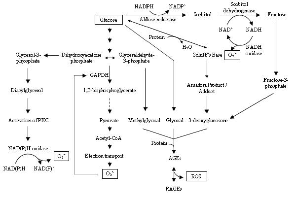
2.3 Formation of AGEs

Surplus production of glyoxal, methylglyoxal and 3-deoxyglucosone from the auto-oxidation of glucose and the fragmentation of glyceraldehyde-3-phosphate (G3P) has been shown to react non-enzymatically with the amino groups of proteins to form cross-links and adducts called AGEs (Brownlee, 1992). The production of AGEs damages target cells by inducing receptor-mediated generation of ROS upon binding with RAGEs and leads to the activation of the nuclear transcription factor, NF-ĸB that mediates pathological changes in diabetic complications (Yan et al., 2003).


2.4 Activation of PKC


Accumulation of G3P due to the inhibition of glyceraldehye-3-phosphate dehydrogenase (GADPH) by mitochondrial O2•- is shown to stimulate the de novo synthesis of diacylglycerol from dihydroxyacetone phosphate, through reduction of the latter to glycerol-3-phosphate and stepwise acylation (Wolf et al., 1991). The diacylglycerol thus formed will activate PKC isoforms that stimulate NAD(P)H-dependent oxidases and lead eventually to the generation of more ROS (Inoguchi et al., 2003).


3. Pathophysiology of Diabetes and Diabetic Complications


There is considerable evidence that the ROS generated during diabetes may activate stress-sensitive signalling pathways that lead to the pathogenesis and pathophysiology of diabetes and diabetic complications. In fact, ROS-mediated insulin resistance, endothelial dysfunctions and β-cell destruction have been reported with diabetic subjects and experimental animals (Evans et al., 2002).


3.1 Free Radicals and Insulin Resistance


Oxidative stress has been shown to activate multiple serine kinase cascades that target both the insulin receptor (IR) and the insulin receptor substrate (IRS) family of proteins. Increased phosphorylation of IR and IRS on discrete serine or threonine residues decreases the extent of insulin-stimulated tyrosine phosphorylation and leads eventually to the impairment of insulin action (Paz et al., 1997). Furthermore, the serine/threonine phosphorylated forms of IRS are less able to associate with IR and downstream target molecules, and result overall, in insulin resistance (Birnbaum, 2001).


3.2 Free Radicals and Endothelial Dysfunctions


Superoxide overproduction has been shown to stimulate the expression of inducible nitric oxide synthase that leads to an overall surplus production of nitric oxide. This in turn, will be quenched by O2•- to form the stronger oxidant, peroxynitrite that is capable of initiating DNA single-strand breakage. DNA damage on the other hand, is an obligatory stimulus for the activation of poly (ADP-ribose) polymerase that results in a cascade mechanism leading to acute endothelial dysfunctions (Ceriello, 2003).


3.3 Free Radicals and β-cell Destruction


Autoimmune destruction of pancreatic β-cells typical of type 1 diabetes mellitus is greatly contributed by ROS and other proinflammatory cytokines such as TNF-{alpha}, IL-1ß, and IFN-{gamma}, whose synergistic interaction results in the ultimate apoptosis and necroticism of pancreatic cells (Hoorens et al., 2001). This is due to the fact that β-cells contain very low free radical-scavenging enzymes that render them more vulnerable to ROS-mediated oxidative damage (Tiedge et al., 1997). On the other hand, transgenic mice with β-cell-targeted overexpression of Cu/Zn-superoxide dismutase, are resistant to alloxan-induced diabetes, thus providing direct in vivo evidence that ROS metabolism can affect susceptibility to oxidative stress-mediated diabetogenesis (Kubisch et al., 1997).


4. Biomarkers of Oxidative Stress


Direct measurement of ROS as biomarkers of oxidative stress is made complicated due to their high reactivity and short half-lives (Jakus, 2000). Nevertheless, the low chemical specificity of ROS that renders them highly damaging to cellular macromolecules, is worth an indirect assessment of oxidative stress. This method evaluates not only levels of damaged biological products, but also reflects an overall antioxidant status of a system.


4.1 Lipid peroxidation


Lipid peroxidation is commonly assessed as an in vivo marker of oxidative stress. Malondialdehyde (MDA) produced from the peroxidation of polyunsaturated fatty acids (Esterbauer et al., 1991) and F2-isoprostanes from free-radical oxidation of arachidonate containing-phospholipids (Gopaul et al., 1994) are the usual measures of cellular lipid peroxidation.

4.2 Oxidation of proteins


Primary products of protein oxidation from the direct reaction of proteins with ROS often leads to accompanying changes in the structures of amino acids such as o-tyrosine, nitrotyrosine, methionine sulfoxide and oxohistidine. Secondary oxidative damage of proteins in contrast, results from the reaction of proteins with reactive carbonyl compounds that arise from the oxidation products of lipids, carbohydrates and proteins. MDA adducts of lysine, carboxymethyllysine and pentosidine are typical examples of such damage (Baynes and Thorpe, 1999).


4.3 Oxidation of nucleic acids


Attack of different reactive species on DNA may be distinguished from the pattern of damage inflicted to the bases. For instance, O2•- and H2O2 are quite latent towards DNA bases while •OH produces a multiplicity of products from all the four bases. In fact, guanine is the most oxidisable base in the DNA and an increase in the level of its oxidation product, 8-hydroxy-2’-deoxyguanosine in the urine of diabetic patients is an indication of oxidative damage (Leinonen et al., 1997).


5. Antioxidant networks


Antioxidants entail their protective mechanisms against oxidative damage by scavenging free radicals or interrupting radical chain reaction. Their protective roles involve a series of network of antioxidants that are strategically compartmentalised in subcellular organelles within the cell to provide maximum protection. For instance, both the Mn- and Cu/Zn-superoxide dismutase remove O2•- by catalysing a dismutation reaction to H2O2 in the mitochondria and cytoplasm, respectively. The reactive H2O2 will be further removed by catalase in the peroxisome or coupled to the oxidization of GSH into GSSG by the mitochondrial and cytosolic glutathione peroxidase (Yu, 1994).


However, the antioxidant defence system is often breached in a diseased state and rapid decline from the baseline level has been noted in animal models with diabetic history compared to normal rats (West, 2000). Nevertheless vitamin E supplementation has been shown to protect LDL against oxidation, which is a critical step in the development of atherosclerosis in late diabetes mellitus. In addition, this vitamin has also been experimentally indicated to improve significantly diabetes-induced abnormal contractility and endothelial dysfunction in diabetes mellitus (Karasu et al., 1997). Vitamin C on the other hand, scavenges ROS in the interstitial fluids and bloodstream and acts to regenerate vitamin E and to increase cellular GSH (Stall and Sies, 1997).

Alpha-lipoic acid in contrast, exists in both the oxidised and reduced forms and acts as an antioxidant in both the membrane and aqueous phase. It establishes cellular antioxidant network by raising intracellular GSH levels and regenerating vitamins E and C. Apart from that, it has also been shown to prevent diabetic retinopathy, alleviates cataract and inhibits aldose reductase activity (Nawroth et al., 2000). In fact, the supplementation of antioxidants and phytochemicals from dietary sources may help to alleviate and delay diabetes induced-oxidative stress.


6. References


Bakker, S.J.L., Ijzerman, R.G., Teerlink, T., Westerhoff, H.V., Gans, R.O.B. and Heine, R.J. 2000. Cytosolic triglycerides and oxidative stress in central obesity: the missing link between excessive atherosclerosis, endothelial dysfunction and β-cell failure? Atherosclerosis 148: 17-21.

Baynes, J.W. and Thorpe, S.R. 1999. Role of oxidative stress in diabetic complications. A new perspective on an old paradigm. Diabetes 48: 1-9.

Birnbaum, M.J. 2001. Turning down insulin signaling. The Journal of Clinical Investigation 108(5): 655-659.

Bradford, M.M. 1976. A rapid and sensitive method for the quantification of microgram quantities of protein utilizing the principle of protein-dye binding. Analytical Biochemistry 72: 248-254.

Brownlee, M. 1992. Glycation products and the pathogenesis of diabetic complications. Diabetes Care 15(12): 1835-1843.

Brownlee, M. 2001. Biochemistry and molecular cell biology of diabetic complications. Nature 414: 813-820.

Ceriello, A. 2003. New insights on oxidative stress and diabetic complications may lead to a “causal” antioxidant therapy. Diabetes Care 26(5): 1589-1596.

Chung, S.S.M., Ho, E.C.M., Lam, K.S.L. and Chung, S.K. 2003. Contribution of polyol pathway to diabetes-induced oxidative stress. Journal of the American Society of Nephrology 14: S233-S236.

Dhar, P., Ghosh, S. and Bhattacharyya, D.K. 1999. Dietary effects of conjugated octadecatrienoic fatty acid (9 cis, 11 trans, 13 trans) levels on blood lipids and nonenzymatic in vitro lipid peroxidation in rats. Lipids 34:109-114.

Esterbauer, H., Schaur, R.J. and Zollner, H. 1991. Chemistry and biochemistry of 4-hydroxynonenal, malonaldehyde and related aldehydes. Free Radical Biology and Medicine 11: 81-128.

Evans, J.L., Goldfine, I.D., Maddux, B.A. and Grodsky, G.M. 2002. Oxidative stress and stress-activated signaling pathways: a unifying hypothesis of type 2 diabetes. Endocrine Reviews 23(5): 599-622.

Flohé, L. and Günzler, W.A. 1984. Assays of glutathione peroxidase. Methods in Enzymology 105: 114-120.

Ganguly, C., De, S. and Das, S. 2000. Prevention of carcinogen-induced mouse skin papilloma by whole fruit aqueous extract of Momordica charantia. European Journal of Cancer Prevention 9(4): 283-288.

Gopaul, N.K., Nourooz-Zadeh, J., Mallet, A.I. and Anggard, E.E. 1994. Formation of F2-isoprostanes during aortic endothelial cell-mediated oxidation of low density lipoprotein. FEBS Letters 348: 297-300.

Grover, J.K. and Yadav, S.P. 2004. Pharmacological actions and potential uses of Momordica charantia: a review. Journal of Ethnopharmacology 93: 123-132.

Higashino, H., Suzuki, A., Tanaka, Y. and Pootakham, K. 1992. Hypoglycemic effects of Siamese Momordica charantia and Phyllanthus urinaria extracts in streptozotocin-induced diabetic rats (the 1st report). Nippon Yakurigaku Zasshi 100(5): 415-421.

Hoorens, A., Stangé, G., Pavlovic, D. and Pipeleers, D. 2001. Distinction between interleukin-1-induced necrosis and apoptosis of islet cells. Diabetes 50: 551-557.

Inoguchi, T., Sonta, T., Tsubouchi, H., Etoh, T., Kakimoto, M., Sonoda, N., Sato, N., Sekiguchi, N., Kobayashi, K., Sumimoto, H., Utsumi, H. and Nawata, H. 2003. Protein kinase C-dependent increase in reactive oxygen species (ROS) production in vascular tissues of diabetes: role of vascular NAD(P)H oxidase. Journal of the American Society of Nephrology 14: S227-S232.

Jacob, S., Lehmann, R., Rett, K. and Häring, H-U. 2000. Oxidative stress and insulin action: a role for antioxidants? In Antioxidants in diabetes management, ed. L. Packer, P. Rösen, H.J. Tritschler, G.L. King and A. Azzi, pp185-204. New York: Marcel Dekker, Inc.

Jakus, V. 2000. The role of free radicals, oxidative stress and antioxidant systems in diabetic vascular disease. Bratisl Lek Listy 101(10): 541-551.

Karasu, C., Ozansoy, G., Bozkurt, O., Erdogan, D. and Omeroglu, S. 1997. Antioxidant and triglyceride-lowering effects of vitamin E associated with the prevention of abnormalities in the reactivity and morphology of aorta from streptozotocin-diabetic rats. Antioxidants in Diabetes-Induced Complications (ADIC) stuffy group. Metabolism 46: 872-879.

Korshunov, S.S., Skulachev, V.P. and Starkov, A.A. 1997. High protonic potential actuates a mechanism of production of reactive oxygen species in mitochondria. FEBS Letters 416: 15-18.

Kubisch, H.M., Wang, J., Bray, T.M. and Phillips, J.P. 1997. Targeted overexpression of Cu/Zn superoxide dismutase protects pancreatic beta-cells against oxidative stress. Diabetes 46(10): 1563-1566.

Leinonen, J., Lehtimaki, T., Toyokuni, S., Okada, K., Tanaka, T., Hiai, H., Ochi, H., Laippala, P., Rantalaiho, V., Wirta, O., Pasternack, A. and Alho, H. 1997. New biomarker evidence of oxidative DNA damage in patients with non-insulin-dependent diabetes mellitus. FEBS Letters 417: 150-152.

Matsuda, H., Li, Y., Murakami, T., Matsumura, N., Yamahara, J. and Yoshikawa, M. 1998. Antidiabetic principles of natural medicine. III. Structure-related inhibitory activity and action mode of oleanolic acid glycosides on hypoglycemic activity. Chemical and Pharmaceutical Bulletin 46(9): 1399-1403.

Miura, T., Itoh, C., Iwamoto, N., Kato, M., Kawai, M., Park, S.R. and Suzuki, I. 2001. Hypoglycemic activity of the fruit of the Momordica charantia in type 2 diabetic mice. Journal of Nutritional Science and Vitaminology 47(5): 340-344.

Nawroth, P.P., Borcea, V., Bierhaus, A., Joswig, M., Schiekofer, S. and Tritschler, H.J. 2000. Oxidative stress, NF-ĸB activation and late diabetic complications. In Antioxidants in diabetes management, ed. L. Packer, P. Rösen, H.J. Tritschler, G.L. King and A. Azzi, pp185-204. New York: Marcel Dekker, Inc.

Nishikawa, T., Edelstein, D., Du, X.L., Yamagishi, S-I., Matsumura, T., Kaneda, Y., Yorek, M.A., Beebe, D., Oates, P.J., Hammes, H-P., Giardino, I. and Brownlee, M. 2000. Normalizing mitochondrial superoxide production blocks three pathways of hyperglycemic damage. Nature 404: 787-790.

Ohkawa, H., Ohishi, N. and Yagi, K. 1979. Assay for lipid peroxides in animal tissues by thiobarbituric acid reaction. Analytical Biochemistry 95: 351-358.

Paz, K., Hemi, R., LeRoith, D., Karasik, A., Elhanany, E., Kanety, H. and Zick, Y. 1997. A molecular basis for insulin resistance. The Journal of Biological Chemistry 272(47): 29911-29918.

Sarkar, S., Pranava, M. and Marita, A.R. 1996. Demonstration of the hypoglycemic action of Momordica charantia in a validated animal model of diabetes. Pharmacological Research 33(1): 1-4.

Sinha, A.K. 1972. Colorimetric assay of catalase. Analytical Biochemistry 47: 389-394.

Sitasawad, S.L., Shewade, Y. and Bhonde, R. 2000. Role of bitter gourd fruit juice in STZ-induced diabetic state in vivo and in vitro. Journal of Ethnopharmacology 73: 71-79.

Sreejayan, M.N.A.R. 1991. Oxygen free radical scavenging activity of the juice of Momordica charantia fruits. Fitoterapia 62(4): 344-346.

Stall, W. and Sies, H. 1997. Antioxidant defense: vitamins E and C and carotenoids. Diabetes 46(S2): S14-S18.

Swidan, S.Z. and Montgomery, P.A. 1998. Effect of blood glucose concentrations on the development of chronic complications of diabetes mellitus. Pharmacotherapy 18(5): 961-972.

Tiedge, M., Lortz, S., Drinkgern, J. and Lenzen, S. 1997. Relation between antioxidant enzyme gene expression and antioxidative defence status of insulin-producing cells. Diabetes 46: 1733-1742.

Ukeda, H., Maeda, S., Ishii, T. and Sawamura, M. 1997. Spectrophotometric assay for superoxide dismutase based on tetrazolium salt 3’-{1-[(Phenylamino)-carbonyl]-3,4-tetrazolium}-bis(4-methoxy-6-nitro)benzenesulfonic acid hydrate reduction by xanthine-xanthine oxidase. Analytical Biochemistry 251: 206-209.

West, I.C. 2000. Radicals and oxidative stress in diabetes. Diabetic Medicine 17: 171-180.

Wolf, B.A., Williamson, J.R., Easom, R.A., Chang, K., Sherman, W.R. and Turk, J. 1991. Diacylglycerol accumulation and microvascular abnormalities induced by elevated glucose levels. The Journal of Clinical Investigation 87: 31-38.

Yan, S.F., Ramasamy, R., Naka, Y. and Schmidt, A.M. 2003. Glycation, inflammation and RAGE. A scaffold for the macrovascular complications of diabetes and beyond. Circulation Research 93: 1159-1169.

Yu, B.L. 1994. Cellular defenses against damage from reactive oxygen species. Physiological Reviews 74(1): 139-162.

No comments: